Healthy Stem Cell Services
In vitro assessment of the effects on the vertebrate genome is currently based on conventional, destructive nuclear staining procedures applied to immortalized mammalian cell lines (OECD TG 487). The use of our self-signaling cell line KCBGFP (DSMC ACC3285) has an excellent potential to replace the previously used lines. Our technique to visualize nuclear structures based on inherited green fluorescent labeling of functional chromosomes reduces the cost of the method on the order of more than 80%.
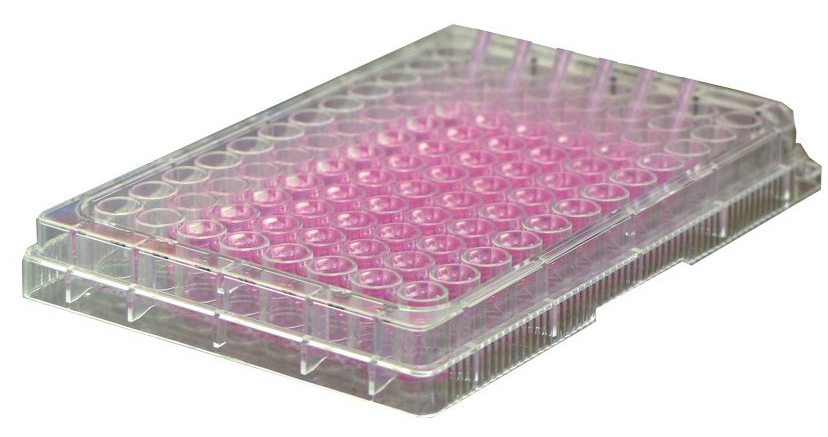
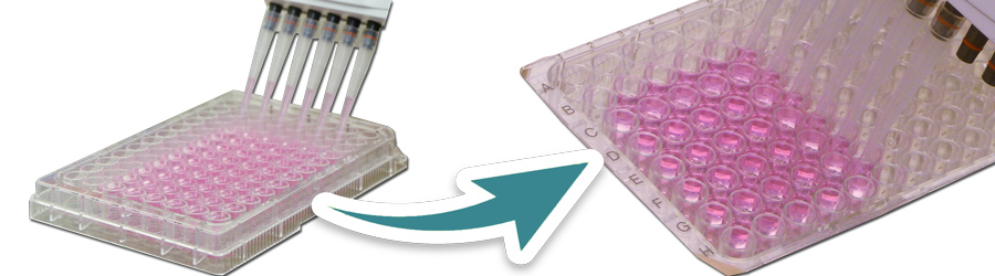
Cell coated plate technology
This new technology enables providing ready-to-use cell coated 96-well culture plates.
After a short pre-culture period (12-24 hrs) under ambient temperature and CO2 conditions the cells grow to a 60% confluent monolayer and are then ready for exposure.
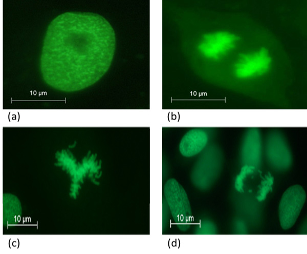
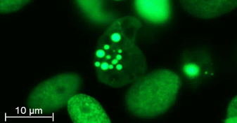
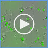
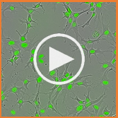
Self-signaling nuclear structures of healthy cells (a-b)
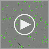
Loss of chromosomes caused by genotoxic compounds (c-d)
Exposure to Test compound
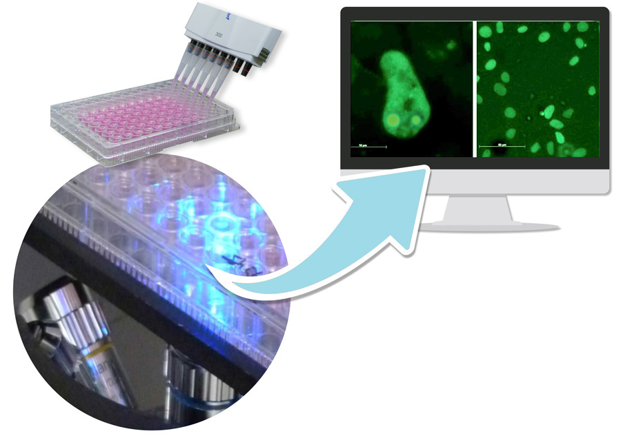
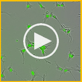
Effects on nuclear structures of living cells can be recorded after 12-14 h exposure to the test compound and a recovery period of 2-24 hours by inverse fluorescence microcopy.
Genotoxicity, Cytotoxicity and Cytostatic Testing
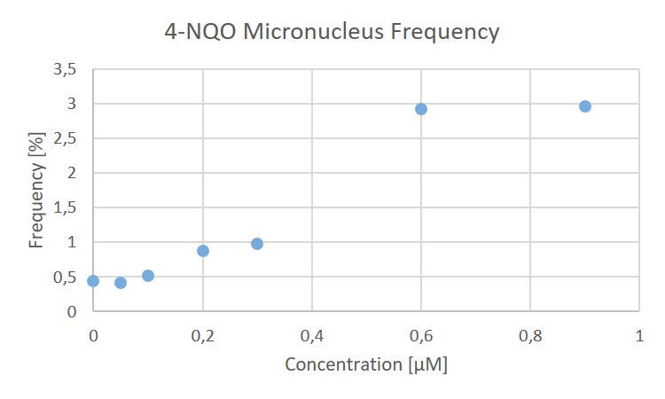
The KCBeGFP cell line allows for the detection of micronuclei. Micronucleus frequencies can be determined in a time window of 2-24h after exposure to the test compound in the living cells.
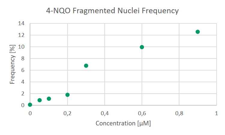
The fragmentation of nuclei indicating cell death due to exposure to a cytotoxic substance is clearly recognizable in pictures of the KCBeGFP cell line. Cytotoxicity can be detected much earlier than previously possible. Brightfield microscopic detectable cell damages occur much later.
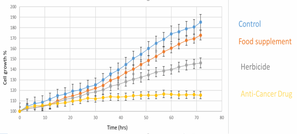
Through kinetic live-imaging of the KCBeGFP cell line, cytotoxic effects can be distinguished from cytostatic effects, an often neglected difference. This enables testing of compounds for non-cytotoxic cytostatic activity. In cancer research drugs that act on cell cycle control provide important information and open new perspectives for therapeutical use.
Downstream analysis
Since the new technique of detecting genotoxins is non-destructive, and the KBCGFP line is neither immortalised nor of malignant origin, a so called “downstream analysis” is possible. After the assessment of the genotoxic potential, further cultivation and investigation regarding carcinogenic transformations of the daughter cells can be performed.
Carcinogenic Cell Transformation
Cell proliferation
The nuclear fluorescence signal enables automated cell counting. The precise reproduction of the chromosomal structure in the course of the cell cycle allows for a distinction between interphase, metaphase, anaphase and cell death (karyorrhexis). The synopsis of these data results in a more differentiated picture of cell proliferation than has been possible so far.
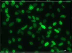
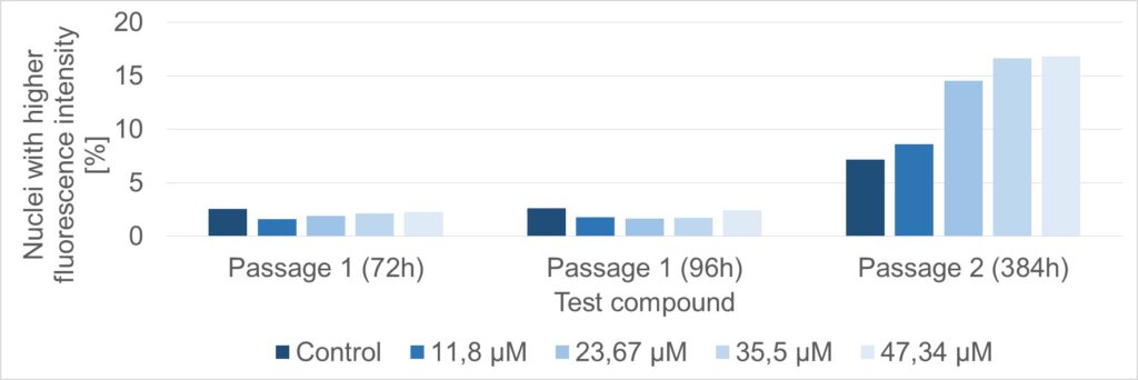
Transformation
After the assessment of the genotoxic potential, further cultivation and investigation regarding carcinogenic transformations of the daughter cells can be performed. The figure shows cells of a down stream culture 384 hours after exposure and subsequent removal of the test compound. The number of nuclei with a brighter fluorescence signal is higher in exposed cells compared to control cells. These alterations of the nuclear morphology correspond to the irreversible condensation of chromatin, known as pyknosis, which is thought to be associated with the carcinogenic transformation of stem cells.
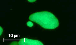
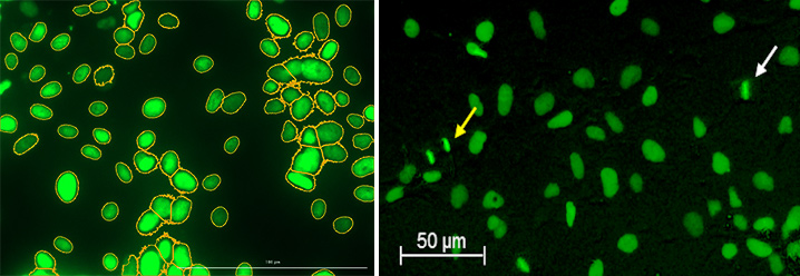
Motility
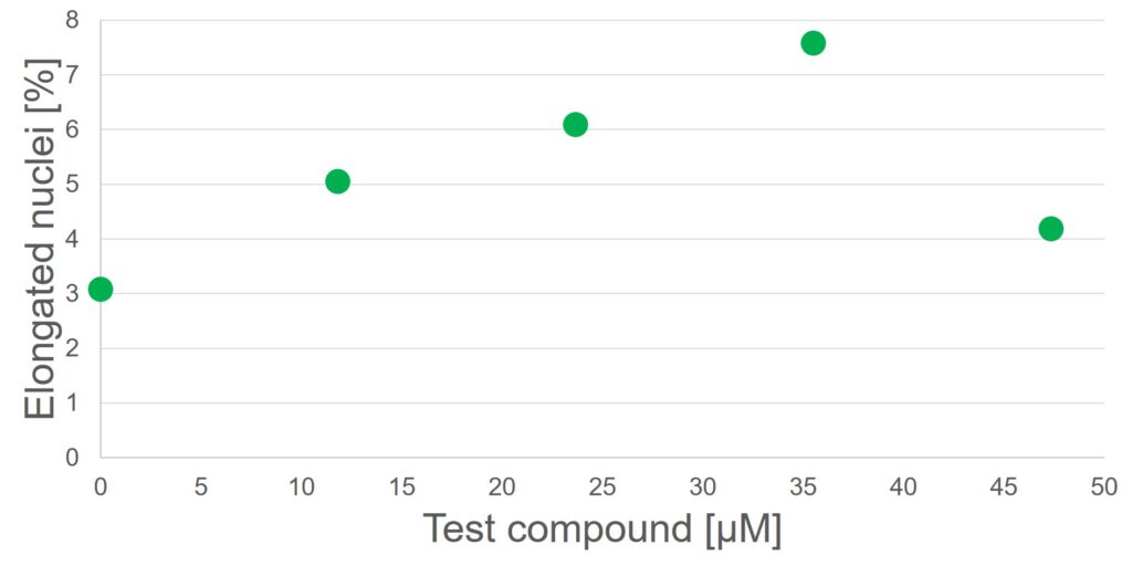
In general in vitro toxicology as well as cytopathology records only snapshots of cell cultures status or the condition of a tissue. Thus role of cell motility is often not considered. Cell boundaries of moving cells in culture or in a tissue are hardly depictable. The elongated deformation of cell nuclei coming along with cell motility, is traceable by the fluorescence signal. For some test compounds a dose-dependent increase in the number of elongated deformed nuclei was observed.
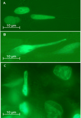
A), B) deformed nuclei of migrating cells in vitro
C) Infiltrating KCB cells in liver ex vivo preparations seven days after transplantation
Molecular Analyses
Gobio has investigated a number of tumorgenes. Please contact us regarding molecular downsstream analyses of post exposure (down stream cells) to investigate marker genes in the focus of your research NGS data of transcriptom available